


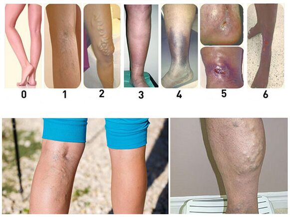
Varicose veins (varicose veins) are diseases in which the superficial veins enlarge or swell. The disease in most cases occurs in people over 30 years of age. In most cases, it is observed on the lower part of the foot. Varicose veins are characterized by the expansion of the venous lumen with simultaneous changes in its wall. The saphenous vein is well contoured, the direction of its travel being "serpentine". Large saphenous veins are usually affected, less frequently small saphenous veins, and more rarely their saphenous anastomosis.
The cause of varicose veins
Theories proposed to explain the causes and mechanisms of disease onset can be reduced to three groups.
The first group theory explains the origin of varicose veins by the anatomical features of the location and structure of these lower limb vessels. The veins have valves that block the centrifugal flow of blood and thus excess flow from the subcutaneous into the veins in the legs. With a lack of valves in the saphenous veins, more blood is deposited, which leads to their expansion.
The theory of the second group in the development of varicose veins emphasizes blood stasis in the pelvis during pregnancy, constipation, as a result of inflammatory processes, as well as during prolonged stay in the legs.
The theory of the third group, which explains the origin of varicose veins by a constitutional tendency, mesenchymal weakness, is the most unfounded.
With varicose veins, for various reasons, their walls change, become thinner, so the increase in pressure leads to bulging of the walls. It first manifests itself in the form of nodes, and at the same time, areas of compaction due to excessive growth of connective tissue are also observed. Mechanical factors only contribute to the development of pathological processes in the veins, but in no way are the main points of pathogenesis, etiology and causes of varicose veins in the lower extremities.
Symptoms of varicose veins
With varicose veins, patients usually experience a feeling of fullness and heaviness in the lower part of the leg. Sometimes there is a nature of short-term pain and seizures. Often there is swelling. The feeling of fullness and heaviness in the limbs increases in the evening, as the edema usually increases during this time. Itching appears, there is often scratching on the feet. In the later stages of the disease, ulcers form, usually located in the lower third of the lower leg on the inside.
The main objective symptom of this disease is visible varicose veins. Examination of the patient to identify these symptoms is performed in a standing position. At the same time, the dilated saphenous vein is clearly visible; on the lower legs they appear more prominent, more convoluted; in the thigh, the vein is usually dilated only along the course of the main vascular trunk. Occasionally there is a varicose vein in the thigh almost at the meeting of the largest saphenous vein into the femoral vein. Such a node can be mistaken for a femoral hernia, but the softness of the node, rapid filling with blood after taking the examiner’s hand and the presence of dilated veins in the lower leg provide the basis for establishing a correct diagnosis.
There are several symptoms that indicate the presence of expansion of the large saphenous vein trunk. These include symptoms where the patient is placed in a horizontal position, the legs are given an elevated position. By rubbing the foot carefully from the edge to the center, the subcutaneous venous system is emptied, the place where the largest saphenous vein flows into the femoral vein is pressed firmly with the fingers and, holding the fingers, the patient is moved to a standing position. If vein filling occurs only after removal of the finger, then this is a positive symptom. In such cases, the anastomosis between the superficial and deep vein networks is less pronounced, and surgery may have a positive effect. If, in an upright position in the patient, the veins at the periphery begin to fill slowly, this indicates the development of a significant anastomosis - a negative symptom. In this case, the vein ligation operation will not be successful.
Delbe-Perthes symptoms show how clearly the saphenous vein empties into the deep through the anastomosis. Elastic bandages are applied to patients in a standing position at the border of the middle and third lower thighs, then they are offered to walk a little. If the tension of the dilated vein is significantly reduced, this indicates the presence of an anastomosis developed between the superficial and deep veins.
Other symptoms of varicose veins include swelling, eczema skin changes, and ulcers. The swelling is different - from a slight tingling to a noticeable edema, when the skin loses its normal pattern and looks shiny, the girth of the lower leg increases significantly. From the manifestations of eczema, dryness, flaking and, finally, eczema rash are observed. The skin on the bottom of the foot is usually affected. These changes occur as a result of trophic disorders.
Prevention and treatment of varicose veins
Prevention of varicose veins is reduced to a change of profession, if it is associated with prolonged standing, taking steps to defecate regularly, wrapping the legs with elastic bandages or wearing elastic stockings. Bandaging the legs or wearing socks must be done while lying down. For a few minutes, the legs are kept in an elevated position and, only after making sure that the veins are empty, they apply a bandage or wear stockings. The bandage is first applied from the bottom and straight up, avoiding any stretching and squeezing that causes stagnation.
There are several methods for surgical treatment. The operation of ligation of the large saphenous vein in the Scarpov triangle where it flows into the femoral vein is palliative. After this operation, relapses are often observed. Therefore, it is only used in combination with other surgical interventions.
During Bebcock’s surgery, a skin incision is made at the lower end of the large dilated saphenous vein, it is separated and tied. On top of the bandage, it is opened and a long abdominal probe is inserted into the lumen. A second small skin incision is made over the upper end of the dilated vein. The middle end is tied and crossed, below the junction of the veins is tied tightly above the probe, after which it is carefully removed through the lower incision. At the same time, the probe pulls on a vein that has been twisted inwards by the intima. The disadvantage of this method is that hematomas form at the site of the torn anastomosis.
During Madelung’s operation, the dilated vein was cut along. Of all the operations, this intervention is the most radical, providing the best long -term results.
Complications of varicose veins
The most common and most difficult complication of varicose veins to treat is varicose ulcers. These ulcers usually occur in the elderly. They are located on the inside, less frequently on the outside, the lower third surface of the lower leg. These ulcers are the result of chronic malnutrition. They are usually deep, have a necrotic, foul -smelling bottom, and a high callus margin. Ulcers can reach large sizes, encircling the entire lower leg. The skin around it is pigmented, sometimes inflamed, with eczema irritation.
Varicose ulcers should be distinguished from syphilis. Syphilis ulcers are usually located in the upper third of the lower leg, more often on the anterior surface. In addition, with syphilis ulcers, other signs of syphilis can be detected. Skin tuberculosis (lupus) is more common on the face, more rarely on the legs. Lupus begins as an isolated nodule that later becomes an ulcer; in the future, deeper tissue damage occurs, sometimes with the formation of smooth scars that tighten neighboring tissues.
Since varicose ulcers develop against the background of circulatory and trophic disorders, their treatment must be continuous and lengthy. The constant position of the patient with the leg raised in most cases leads to a rapid improvement. Bandages with a solution of 0. 5% potassium permanganate, with penicillin ointment or balsamic liniment should be placed on the ulcer. When the wound is cleaned and the swelling around it disappears, it is recommended to remove the vein. Only radical surgery to remove the altered vein can eliminate the risk of recurrent ulcers.
As the disease progresses and the varicose nodes increase, the walls and skin soldered to it become thinner. As a result, usually during walking (when the nodes are very tense), one of the nodes may rupture and venous bleeding may occur. Although such bleeding can be significant, it does not pose a great danger, as it quickly stops if the patient is laid down and the leg is lifted. In this position, negative pressure is created in the veins, they subside and the bleeding stops. A light aseptic bandage is placed on the wound. Due to the fact that bleeding can recur, surgery is recommended for the isolation of the veins or their ligation and the removal of the thinnest nodes. With bleeding from a compensatory dilated vein, any operation associated with the ligation of the main trunk of the vein is categorically contraindicated.