Many people mistakenly believe that varicose veins are just a threat to the appearance of the legs. In fact, everything is more serious-the disease is quite often complicated by thrombosis and inflammation of the deep veins, and in further cases, chronic venous insufficiency, which is indicated by significant trophic changes in the tissues. Therefore, it is necessary to diagnose this pathology at an early stage to prevent the development of dangerous conditions.

Varicose veins are pathological changes in the walls of venous vessels, which occur under the influence of blood accumulated in them. Often, this process develops in the veins of the legs and small pelvis. Usually, blood through the veins is directed only to the heart, this is facilitated by the valves and muscles of the veins, which, by their contraction, seem to "drive" the blood through the vessels. With varicose veins, for some reason, abnormal blood flow is formed. It begins to stagnate first in the deep veins, and then on the superficial ones, which increase, forming varicose veins under the skin.
Symptoms of varicose veins in the lower part of the legs
The first signs of the disease are not specific (they are also found in other diseases), they are combined together under the term "heavy foot syndrome". It is characterized by increased and progressive fatigue in the lower legs, pain in the legs, a feeling of heaviness, burning and rupture in the calves, night cramps of the calf muscles. These symptoms appear at the end of the day, especially if a person has been standing or sitting for a long time during that time. Subsequently, with the development of pathology, evening swelling of the back of the foot and ankle is added to the manifestations of the described disease. After resting, the condition of the sore foot usually improves.
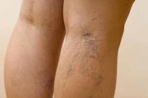
Visual changes in the early stages of the disease are not always noticeable, because varicose veins in the legs begin with deeper channels. The only external sign of a problem that has started may be the vascular network. They, of course, do not always show varicose veins, but it is better to consult a phlebologist, a specialist in varicose veins, when they appear.

But in the final stages of varicose veins, cyanotic subcutaneous veins and varicose nodes have already appeared - these are shallow veins that are enlarged and winding that resemble grapes. They are usually located on the inside of the lower legs and thighs.
In addition, with the development of pathology, the feet begin to swell more. Gradually, chronic venous insufficiency forms, in which venous outflow and microcirculation in the tissues are disrupted. All this is reflected in the condition of the skin of the feet: it becomes dark, flaky, itchy, then a trophic ulcer appears on it, which heals very poorly. This is how varicose veins develop. The same results from varicose veins can be prevented with timely treatment, therefore, if even slight, but systematic discomfort in the legs, and vascular networks or "stars" on the skin appear, you should see a doctor.
Symptoms of pelvic varicose veins
In the pelvis, varicose veins are less common than in the legs and mostly in young women. The trigger for the development of this pathology is pregnancy (both hormonal and mechanical contributing factors play a role here). After childbirth, the symptoms of the disease, as a rule, disappear, and only about 10% of women notice the periodic resumption of unpleasant symptoms after prolonged standing, hypothermia, and physical exertion.
Small pelvic varicose veins are indicated by chronic pelvic pain, as well as the development of superficial vein formation in the perineum and vulva. Such patients often fail to treat inflammatory diseases of the reproductive organs, because of pain in the lower abdomen, characteristic of pelvic varicose veins, sometimes mistakenly associated with chronic oophoritis, salpingitis, endometriosis, etc.
How are varicose veins diagnosed?
When a varicose node becomes clearly visible on a patient’s leg, a doctor can make a diagnosis of "varicose disease" even without the results of an instrumental study. If new pathology begins to develop or is localized in the small pelvis, in -depth examination is essential.

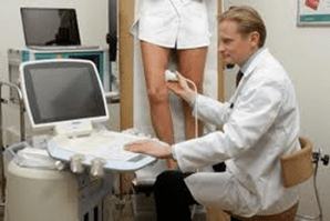
The main method for diagnosing varicose veins is Doppler ultrasound. This study is informative for vein lesions in any part of the body. With the help of ultrasound, the doctor can study the condition of the walls and anatomy of the deep and superficial veins, valves, assess the blood flow in the ducts, detect the backflow of blood, etc. The classification of varicose veins and, thus, the choice of treatment method is based precisely on the results of ultrasound.
Another diagnostic method used in this pathology is rheovasography. Its implementation allows you to determine how well the tissues of the lower legs are filled with blood and with it nutrients. This information helps the doctor to determine at what stage of the disease: at the stage of compensation, subcompensation, etc.
Less often, phlebography is used for varicose veins - this is an X -ray examination of the veins using contrast.
In addition, a comprehensive examination of patients with varicose veins usually includes a variety of blood tests: the doctor is particularly interested in the levels of hemoglobin, erythrocytes, platelets, and coagulogram parameters. These data make it possible to assess the density of blood and the tendency of the patient's body to form blood clots.


